
All About Long-COVID

Please note that this article summarizes research that may be of interest to healthcare professionals and educated laymen but is not intended as any sort of prescription or medical advice. All readers are encouraged to seek a trusted healthcare professional for medical advice.
What Is Long-COVID Syndrome?
Long-COVID Syndrome
Also known as: Long-haul COVID Syndrome
Or:
PASC (Post-acute sequelae of SARS-CoV-2 infection),
is a persistence of the symptoms of Sars-CoV-2 infection past a period of four weeks.
How Does It Manifest?
· Shortness of Breath or Difficult Breathing
· Pronounced Fatigue (Similar to That Experienced in Chronic Fatigue Syndrome)
· Joint Pain
· Memory or Concentration Problems
· Sleep Problems
· Muscle Pain
· Headache
· Loss of Smell (Anosmia) and/or Taste (Aguesia)
· Fast or Pounding heartbeat
· Orthostatic Hypotension
· Depression and/or Anxiety
· Inflammation (Heart or Lungs Most Common)
· Sensory Neuropathies, including Numbness and
Weakness in the Legs
· Worsened Symptoms after Physical or Mental Activities
Who Does It Affect?
Ø Affects 1.3–30% of individuals infected with SARS-CoV-2. (Note that this range is in dispute).
Ø However, 50% of hospitalized COVID patients tend to experience it. In fact, the sicker someone is with the acute infection, and the longer the duration of that infection, the more likely they are to experience long-COVID Syndrome.
Ø This is why early treatment—downplayed by public health officials—is so very, very important! (Data from a group of 3,000 patients treated by Dr. Vladimir Zelenko within the first five days of the onset of COVID-19 showed that none of these went on to develop long-haul symptoms, including fatigue, brain fog, or difficulty breathing.)
Why Does It Affect Them?
A number of possible contributing factors to the etiology of long-haul COVID have been proposed:
Ø 1. Residual organ damage (esp. cardiovascular, pulmonary, and neurological) due to inflammation, possibly instigated by…
Ø 2. Viral persistence (in particular sites within the body which are difficult for the immune system to eradicate--most likely owing to delayed treatment in early stage of SARS Cov-2 infection) and/or immune dysregulation/autoimmunity. (SARS-CoV-2 may disturb self-tolerance and trigger autoimmune responses through cross-reactivity with host cells.)
Ø 3. Depletion of glutathione, the body’s master detoxifier (via the liver’s Phase-Two detox process) and immunosupportive tripeptide. If we’re deficient in it, we’re more likely to get sick and also to experience a perpetuated immune response (above).
Ø 4. Dysbiosis, causing immune imbalances and neuroinflammation (more on this below).
Ø 5. Nutrient deficiencies (C; D; Se; Zn)
Evidence for These Etiological Factors
1. Remaining Virus, Altered Immune Response, & Inflammation
Ø Dr. Bruce Patterson, speaking at the International COVID Summit in Rome in Sept. 2021, shared his research that in “individuals who’ve had significant COVID illness, 15 months later the s1 segment of the spike protein is recoverable from human monocytes.” In a paper, Patterson and colleagues went into great detail, highlighting the “immune response to persistent viral antigens, specifically the S1 fragment of the spike protein” in long-COVID and its association with inflammation, especially vascular inflammation.
Patterson, B. et al. 2022. “Persistence of SARS CoV-2 S1 Protein in CD16+ Monocytes in Post-Acute Sequelae of COVID-19 (PASC) up to 15 Months Post-Infection,” Front Immunol. 12:746021 https://www.frontiersin.org/articles/10.3389/fimmu.2021.746021/full#B3
Additional Studies:
Chertow, D. et al. 2021.“SARS-CoV-2 infection and persistence throughout the human body and brain,” Res Sq. Preprint posted online December 20, 2021.
Proal, A., VanElzakker, M. 2021. “Long COVID or Post-acute Sequelae of COVID-19 (PASC): An Overview of Biological Factors That May Contribute to Persistent Symptoms,” Front Microbiol. 2021 12:698169.
Visvabharathy L. et al. 2021. “Neuro-COVID long-haulers exhibit broad dysfunction in T cell memory generation and responses to vaccination,” medRxiv [Preprint]. Oct 29:2021.08.08.21261763
Peluso, M. et al. 2021. “Markers of Immune Activation and Inflammation in Individuals With Postacute Sequelae of Severe Acute Respiratory Syndrome Coronavirus 2 Infection.” J Infect Dis. 224(11):1839-1848.
Ø A complete blood count (CBC) often shows a skewed ratio of white blood cells, marked by a depressed lymphocyte count. (Lymphocytes, particularly T-cells, participate in inflammation resolution following infection; long-COVID, as we have seen, is marked by chronic inflammation.) Total white blood cells (leukocytes) may also be depressed.
Ø Several studies have found autoantibodies in long-COVID patients, differentiated by sex. One such study:
Liu, Y. et al. 2021. “Paradoxical Sex-specific Patterns of Autoantibody Response to SARS-CoV-2 Infection.” J Trans Med 19:524
Ø Several studies have shown that long-COVID can present as a post-viral conglomeration of different illnesses: chronic fatigue syndrome, mast-cell activation syndrome (MCAS), and dysfunction in the autonomic nervous system. These illnesses can trigger symptoms across every organ system.
Schofield, J. “Persistent antiphospholipid antibodies, mast cell activation syndrome, postural orthostatic tachycardia syndrome and post-COVID syndrome: 1 year on,” Eur J Case Rep Intern Med. 2021;8(3):002378.
Ø A reactivation of other viruses, including Epstein-Barr virus (EBV), cytomegalovirus (CMV), and shingles may occur with COVID. In 2021, several studies demonstrated that Epstein-Barr Virus (EBV) was reactivating in long-haulers and was most likely responsible for their fatigue.
Gold, J. et al. 2021. “Investigation of long COVID prevalence and its relationship to Epstein-Barr virus reactivation,” Pathogens. 10(6):763.
Drago, F. et al. 2021. “Human herpesvirus-6, -7, and Epstein-Barr virus reactivation in pityriasis roseaduring COVID-19. J Med Virol. 93(4):1850-1851.
2. Depleted Glutathione
Researchers at Baylor College of Medicine found that patients hospitalized with COVID-19 had elevated oxidative stress and profoundly reduced levels of glutathione, and that this was true in all age levels, whereas normally such affectations only occur in folks 60+ years of age.
Kumar, P. et al. 2021. “Severe Glutathione Deficiency, Oxidative Stress and Oxidant Damage in Adults Hospitalized with COVID-19: Implications for GlyNAC (Glycine and N-Acetylcysteine) Supplementation,” Antioxidants (Basel). 11(1):50.
Glutathione is crucial for cytokine production and immune response.
Wu, G. et al. 2004. “Glutathione Metabolism and its Implications for Health.” J Nutr. 134(3):489-92; Dröge, Wulf, and Raoul Breitkreutz. 2000. “Glutathione and immune function.” Proceedings of the Nutrition Society 59(4): 595-600
Pronounced fatigue can occur with low levels of glutathione.
3. Residual Organ (CV, Brain, Lung) Damage
When 10 long-COVID patients were examined with an exercise challenge, this resulted in a marked reduced peak exercise aerobic capacity (oxygen consumption [VO2]) along with an exaggerated hyperventilatory response.
Singh, I. et al. 2022. “Persistent Exertional Intolerance After COVID-19: Insights From Invasive Cardiopulmonary Exercise Testing,” Chest. 161(1):54-63.
When 9 long-COVID patients were examined with a variety of tests, the results revealed: cerebrovascular dysregulation with persistent cerebral arteriolar vasoconstriction; small fiber neuropathy and related dysautonomia; respiratory dysregulation; and chronic inflammation.
Novak, P. et al. 2022. “Multisystem Involvement in Post-Acute Sequelae of Coronavirus Disease 19,” Ann Neurol. 91(3):367-379.
Salk Institute researchers created a “pseudovirus” that was surrounded by SARS-CoV-2’s crown of spike proteins, but did not contain any actual virus. Exposure to this pseudo-virus in lab animals damaged their lungs and arteries, inflaming endothelial cells, which proved that the spike protein alone was enough to cause disease. Healthy endothelial cells were then exposed to the spike protein in a Petri dish, which adversely affected their ACE-2 receptors and disrupted these receptors’ signaling to mitochondria, in which damage and fragmentation occurred.
Lei, Y. et al. 2021. “SARS-CoV-2 Spike Protein Impairs Endothelial Function via Downregulation of ACE 2,” Circ Res 128(9):1323-1326.
Persistent micro-clots have been found. These could be blocking capillaries, inhibiting oxygen transport, and entrapping inflammatory molecules.
Pretorius, E. et al. 2021. “Persistent Clotting Protein Pathology in Long COVID/Post-Acute Sequelae of COVID-19 (PASC) Is Accompanied by Increased Levels of Antiplasmin,” Cardiovasc Diabetol 20:172.
Kell, D. et al. 2022. “A central role for amyloid fibrin microclots in long COVID/PASC: origins and therapeutic implications. Biochemical Journal 479:537-59.
Researchers have posited that the presence of spike protein S1 in circulation may contribute to the hypercoagulation in COVID-19 positive patients and may cause severe impairment of fibrinolysis, which may result in the persistent microclots noted in patient plasma samples.
Grobbelaar, L. et al. 2021. “SARS-CoV-2 Spike Protein S1 Induces Fibrin(ogen) Resistant to Fibrinolysis: Implications for Microclot Formation in COVID-19,” Biosci Rep 41 (8): BSR20210611.
See also:
Pretorius, E. et al. 2021. “Persistent clotting protein pathology in Long COVID/Post-Acute Sequelae of COVID-19 (PASC) is accompanied by increased levels of antiplasmin,” Cardiovasc Diabetol 20:172.
Using a newly developed mouse model of acute lung injury, researchers found that exposure to the SARS-CoV-2 spike protein alone was enough to induce COVID-19-like symptoms.
“Sars CoV-2 Spike Protein Alone May Cause Lung Damage” https://medicalxpress.com/news/2021-04-sars-cov-spike-protein-lung.html
See, also, on damage caused by spike protein (S1):
Khaddaj-Mallat, R. et al. 2021. “SARS-CoV-2 Deregulates the Vascular and Immune Functions of Brain Pericytes via Spike Protein.” Neurobiol Dis. 161:105561.
4. Dysbiosis
“Gut microbiome of patients with PACS were characterised by higher levels of Bacteroides vulgatus and lower levels of Faecalibacterium prausnitzii…. Butyrate-producing bacteria, including Bifidobacterium and Faecalibacterium prausnitzii showed the largest inverse correlations with PACS at 6 months.”
Liu, Q, et al. 2022. “Gut microbiota dynamics in a prospective cohort of patients with post-acute COVID-19 syndrome,” Gut. 71(3):544-552.
See also:
Zuo, T. et al. 2020. “Alterations in gut microbiota of patients with COVID-19 during time of hospitalization,” Gastroenterology 159(3): 944–955.e8.
Hazan, S. et al. 2022. “Lost microbes of COVID-19: Bifidobacterium, Faecalibacterium depletion and decreased microbiome diversity associated with SARS-CoV-2 infection severity,” BMJ Open Gastroenterology 9:e000871.
5. Nutrient Deficiencies
Selenium
In one study, 42% of hospitalized COVID patients were deficient in selenium. In another study, 43.3% of COVID-positive patients with fever and dyspnea had low selenium levels compared with 20% of a healthy control group.
Im, J. et al. 2020. “Nutritional status of patients with COVID-19. Int J Infect Dis 100:390-393
Majeed, M. et al. 2021. “An exploratory study of selenium status in healthy individuals and in patients with COVID-19 in a south Indian population: The case for adequate selenium status,” Nutrition. 82:111053.
Butler, M., Barrientos, R. 2020. “The impact of nutrition on COVID-19 susceptibility and long-term consequences,” Brain Behav. Immun. 87:53–54.
Moghaddam A. et al. 2020. “Selenium Deficiency Is Associated with Mortality Risk from COVID-19,” Nutrients. 2020 Jul 16;12(7):2098.
Zinc
One study showed deficiency in 55% of COVID patients.
Jothimani, D, et al. 2020. “COVID-19: Poor outcomes in patients with zinc deficiency,” Int J Infect Dis. 100:343-349.
Vitamin C
Deficient in many COVID-19 sufferers
Arvinte, C. et al. 2020.“Serum levels of vitamin C and vitamin D in a cohort of critically ill COVID-19 patients of a north American community hospital intensive care unit in may 2020: a pilot study,” Med Drug Discov. 8:100064
Patterson, T. et al. 2021. “Low level of Vitamin C and dysregulation of Vitamin C transporter might be involved in the severity of COVID-19 Infection,” Aging and disease. 12(1): 14-26
Vitamin D
Very commonly deficient in COVID-19 sufferers: In one study, 76% of hospitalized COVID patients were deficient in it. In a study of 49 pediatric patients with COVID-19, 82% were deficient.
Im, J. et al. 2020. “Nutritional status of patients with COVID-19. Int J Infect Dis 100:390-393
Arvinte, C. et al. 2020.“Serum levels of vitamin C and vitamin D in a cohort of critically ill COVID-19 patients of a north American community hospital intensive care unit in may 2020: a pilot study,” Med Drug Discov. 8:100064
Karakaya, M. et al. 2021. “Evaluation of nutritional status in pediatric patients diagnosed with Covid-19 infection.’ Clin Nutr ESPEN. 44:424-428.
Labs to Determine Extent of Possible Contributing Factors Referenced Above.
(Many of these Can Be Ordered via Consumer-direct Labs—No Doctor’s Order Required—Such as the Three Labs Listed Below)
www.lifeextension.com/lab-testing (via a local LabCorp)
Persistent Viral Infection / Altered Immune Response / Inflammation / Glutathione level / Blood Clotting
Complete Blood Count (CBC) to Check Lymphocytes, Neutrophils, Hemoglobin, Platelets, etc. Lymphocytes are typically low (= lymphopenia) in severe COVID/long-COVID.
Gamma-Glutamyl Transferase (GGT) (If above 16, indicates that glutathione level is lower than ideal.)
Epstein-Barr Virus panel, consisting of four analytes, most importantly including early antigen (EA), which typically indicates if EBV has recently been reactivated.
AM Cortisol, a marker of immune function, inflammation, and energy levels that is sometimes depressed in long-COVID
Serum IgA, a marker of immune imbalance that is often depressed in long-COVID
C-Reactive Protein, a marker of inflammation
D-Dimer, a marker of blood clotting
Troponin, a type of protein found in heart muscle but not usually in blood. It is released into the blood with cardiac damage. Important to test if there is chest pain and/or tightness and shortness of breath
Serum Ferritin. Often elevated in long-COVID
Serum Creatinine, a marker of kidney damage that is sometimes elevated in long-COVID
Lactobacilli/Bifidobacteria
GI Effects (Genova Diagnostics) or:
GI360™ Microbiome-Doctor's Data Kit
Nutrient Deficiencies
RBC Selenium
Total Vitamin D (25-hydroxy vitamin D)
Vitamin C
RBC Zinc
Therapeutics for Long-COVID
Raise Glutathione Level
· N-acetyl Cysteine (NAC)
Cysteine is a precursor to glutathione. NAC, a form of cysteine, needs to be taken on an empty stomach for best effects, but can cause indigestion in some persons and is contraindicated in ulcers. (Liposomal glutathione can be used as a substitute, but is a lot more expensive.) NAC is best taken with glycine (immediately below).
· Glycine
A precursor to glutathione. Often helpful for sleep, as well, if taken in the evening. Best taken with N-acetyl Cysteine (NAC) (immediately above)
· alpha-Lipoic acid
Can help regenerate glutathione
Zhang J. et al. 2017. "Regeneration of glutathione by α-lipoic acid via Nrf2/ARE signaling pathway alleviates cadmium-induced HepG2 cell toxicity". Environ Toxicol Pharmacol. 51:30–37
· Selenium
Required by glutathione peroxidase to produce glutathione.
· Vitamin B2 (Riboflavin)
Riboflavin is a necessary coenzyme for the activity of glutathione reductase, an enzyme that converts the oxidized glutathione into its reduced form, which is the compound necessary for antioxidant function. (Riboflavin deficiency can cause crusted skin around the nose, cracked skin at the fingernails, genital skin lesions, stomatitis, photophobia, blurred vision, and seborrheic dermatitis.)
· Vitamin C
Helps to spare depletion of glutathione and works with latter to protect mitochondria from oxidative damage.
Meister, A. 1995. “Mitochondrial changes associated with glutathione deficiency.” Biochim Biophys Acta.1271(1):35-42
· Vitamin D
The active metabolite of vitamin D in the body (calcitriol) increases glutathione levels in the brain and appears to be a catalyst for glutathione production. (Oral vitamin D3 can take 10 days to metabolize to calcitriol, however. This conversion depends upon magnesium to activate the enzymes involved.)
Garcion, E. et al. 2020. "New clues about vitamin D functions in the nervous system,” Trends in Endocrinology and Metabolism. 13(3): 100–5
Check Persistent Virus / Support Balanced Immunity / Reduce Inflammatory Response
· n-Acetyl cysteine (NAC)
Antiviral, partly because of its ability to inhibit the protein complex NF-kappa B needed by RNA viruses to replicate. Lymphocytes rely on glutathione to function properly and NAC has been shown to increase the potency of lymphocytes when a virus has depressed them.
Shi, Z., Puyo, C. 2020. “N-Acetylcysteine to Combat COVID-19: An Evidence Review,” Ther Clin Risk Manag. 16:1047-1055.
· Andrographis (Andrographis paniculata)
This Southeast-Asian herb has shown powerful antiviral effects, including against SARS-CoV-2 and Epstein-Barr Virus (EBV)
Sa-Ngiamsuntorn, K. et al. 2021. “Anti-SARS-CoV-2 Activity of Andrographis paniculata Extract and Its Major Component Andrographolide in Human Lung Epithelial Cells and Cytotoxicity Evaluation in Major Organ Cell Representatives,” J Nat Prod. 84(4):1261-1270.
Lin T. et al. 2008. “Inhibition of the epstein-barr virus lytic cycle by andrographolide,” Biol Pharm Bull. 31(11):2018-23.
Malat P. et al. 2021. “Andrographolide Inhibits Lytic Reactivation of Epstein-Barr Virus by Modulating Transcription Factors in Gastric Cancer,” Microorganisms 9(12):2561.
· Black cumin (Nigella sativum) seed oil
CLINICAL TRIAL: “The percentage of recovered [COVID] patients in NSO [Nigella sativa oil] group (54 [62 %]) was significantly higher than in the control group (31[36 %]; p = 0.001). The mean duration to recovery was also shorter for patients receiving NSO (10.7 ± 3.2 days) compared with the control group (12.3 ± 2.8 days); p = 0.001.”—Koshak A. et al. 2021. “Nigella sativa for the treatment of COVID-19: An open-label randomized controlled clinical trial,” Complement Ther Med. 61:102769.
See also:
Esharkawy E. et al. 2022. “In vitro potential antiviral SARS-CoV-19- activity of natural product thymohydroquinone and dithymoquinone from Nigella sativa,” Bioorg Chem. Jan 1;120:105587. Epub ahead of print
· Boneset (Eupatorium perfoliatum)
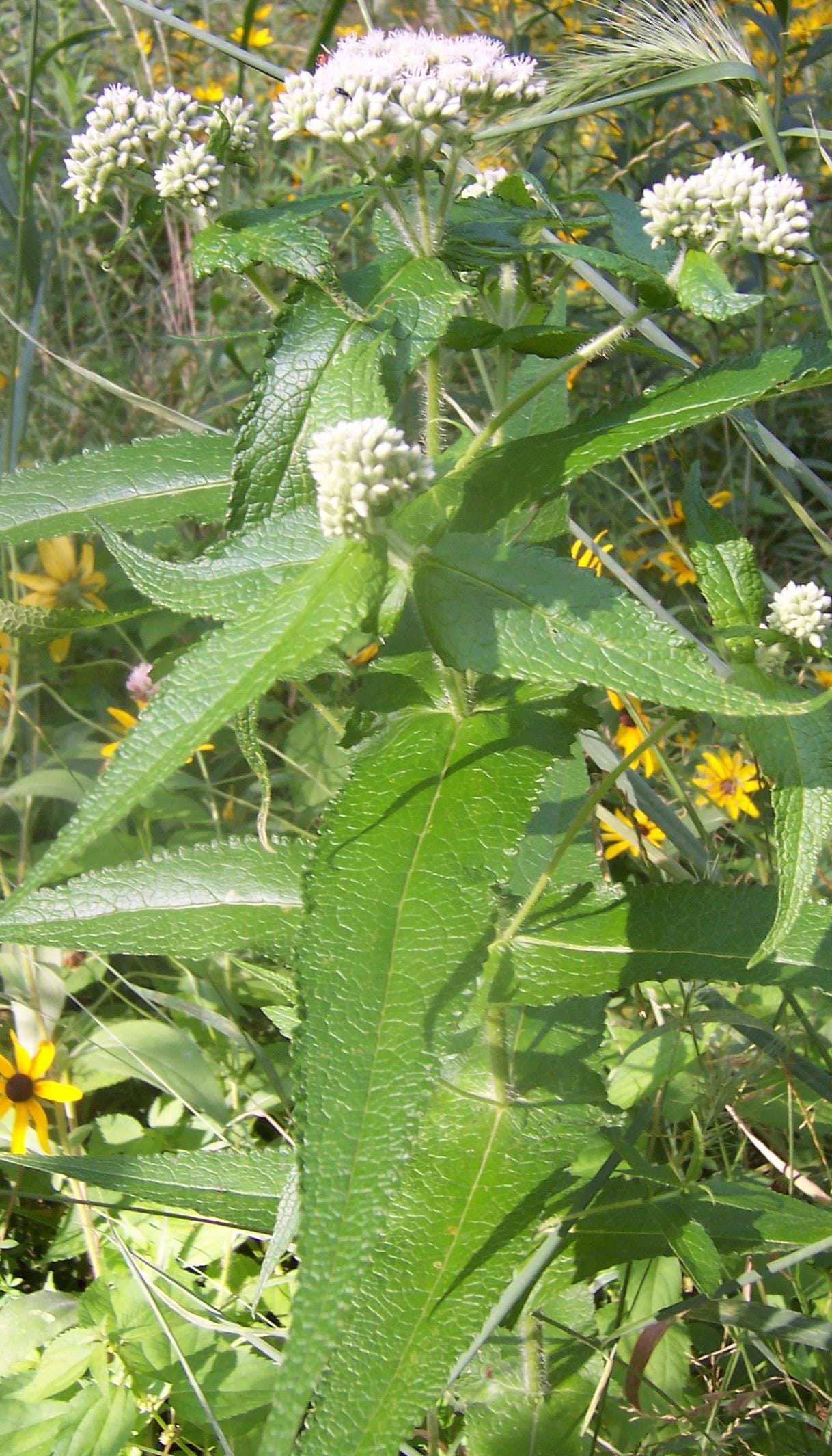
Was used by Eclectic physicians (practicing in the U. S. from the mid-1800s until WW2) for poor immune response and slow recovery from viral infections. Has been shown in scientific studies to be antiviral to both influenza and dengue. Classically used also for aching in the chest with restricted breathing. Also has stimulant properties to overcome viral fatigue.
Sinha M. et al. 2022. “In-vitro antiviral action of Eupatorium perfoliatum against dengue virus infection: Modulation of mTOR signaling and autophagy.” J Ethnopharmacol. 282:114627.
Derksen, A. 2016. “Antiviral activity of hydroalcoholic extract from Eupatorium perfoliatum L. against the attachment of influenza A virus,” J Ethnopharmacol. 188:144-52.
· Cat’s claw [Una de Gato](Uncaria tomentosa)
Two modeling studies and an in vitro study reveal powerful antiviral potential for this herb against SARS-CoV-2. In the in vitro study, it had a 92%+ effect in the SARS-CoV-2 viral titer and CPE [cytopathic effect] after 48 hours of treatment.
Yepes-Perez, A. et al. 2021. “The Hydroalcoholic Extract of Uncaria tomentosa (Cat's Claw) Inhibits the Infection of Severe Acute Respiratory Syndrome Coronavirus 2 (SARS-CoV-2) In Vitro,” Evid Based Complement Alternat Med. 2021 Feb 24;2021:6679761.
· Chinese (Baikal) skullcap (Scutellaria baicalensis)
The ethanol extract of S. baicalensis and its major component, baicalein, have been shown to inhibit SARS-CoV-2 3CLpro activity in vitro. Both of them inhibit the replication of SARS-CoV-2. While baicalein is mainly active at the viral post-entry stage, a tincture of the whole herb inhibits viral entry. Also shown to inhibit Epstein-Barr Virus-EA activation. Baicalein is also a powerful anti-inflammatory and anti-allergic compound
Liu, H. et al. 2021. “Scutellaria baicalensis extract and baicalein inhibit replication of SARS-CoV-2 and its 3C-like protease in vitro,” J Enzyme Inhib Med Chem. 36(1):497-503. doi: 10.1080/14756366.2021.1873977.
Konoshima, T. et al. 1992. “Studies on inhibitors of skin tumor promotion. XI. Inhibitory effects of flavonoids from Scutellaria baicalensis on Epstein-Barr virus activation and their anti-tumor-promoting activities,” Chem Pharm Bull (Tokyo). 40(2):531-3
Dinda, B. et al. 2017. “Therapeutic potentials of baicalin and its aglycone, baicalein against inflammatory disorders,” European journal of medicinal chemistry. 131:68-80.
· Cordyceps (Cordyceps sinensis)

Helpful with immune weakness. One of its major compounds has been shown to suppress EBV replication.
Ryu, E. et al. 2014. “Cordycepin is a novel chemical suppressor of Epstein-Barr virus replication,” Oncoscience. 1(12):866-881.
Ryu, E. et al. 2014. “Cordycepin is a novel chemical suppressor of Epstein-Barr virus replication,” Oncoscience. 1(12):866-881.
· EGCG ( a compound from green tea)
Antiviral to Epstein-Barr Virus (EBV) lytic cycle.
Chang L. et al. 2003. “Inhibition of Epstein-Barr virus lytic cycle by (-)-epigallocatechin gallate. Biochem Biophys Res Commun. 301(4):1062–1068.
Liu, S. et al. 2013. “(-)-Epigallocatechin-3-gallate inhibition of Epstein-Barr virus spontaneous lytic infection involves ERK1/2 and PI3-K/Akt signaling in EBV-positive cells,” Carcinogenesis. 34(3):627–637.
· Iodine
Supports the body’s surveillance system for removing abnormal cells; inactivates viruses by interfering with their protein coat that enables them to adsorb to host cells; and triggers a mechanism for apoptosis (normal programmed death of cells as part of their life cycle) in cells infected with viruses.
Altaf, I. et al. 2021. “An in vitro antiviral activity of iodine complexes against SARS-CoV-2,” Arch Microbiol 203:4743–4749
Kariwa H. et al. 2006. “Inactivation of SARS Coronavirus by Means of Povidone-Iodine, Physical Conditions and Chemical Reagents,” Dermatology 212(suppl 1):119-123.
Verheesen, R., Traksel, R. 2020. “Iodine, a preventive and curative agent in the COVID-19 pandemic?” Medical hypotheses, 144:109860.
· L-Lysine
Crucial supplement for all herpes-family viruses, incl. reactivation of Epstein-Barr virus and of Shingles. Typical dose is 500 mg., b.i.d. to t.i.d. Needs to be taken on an empty stomach. For more on L-lysine’s potental, see:
· Monolaurin
A component of coconut oil that has been clinically shown to be quite effective against many viruses, including EBV. In one study, monolaurin “removed all measurable infectivity from all of the [14 RNA and DNA] viruses tested” and “reduced infectivity by disintegrating the virus envelope.”
Hierholzer. J., Kabara, J. 1982. “In vitro effects of monolaurin compounds on enveloped RNA and DNA viruses,” J Food Saf. 4(1):1-12.
· Moringa (Moringa oleifera)
Constituents have been shown to inhibit EBV early antigen.
Hussain, S. et al 2014. “Review: an exposition of medicinal preponderance of Moringa oleifera (Lank.),”Pakistan Journal of Pharmaceutical Sciences 27(2):397-403
· Motherwort (Leonurus cardiaca)
Contains a high amount of ursolic acid, shown to inhibit EBV
Tokuda, H. et al. 1986. “Inhibitory effects of ursolic and oleanolic acid on skin tumor promotion by 12-O-tetradecanoylphorbol-13-acetate,” Cancer Lett 33: 279–285.
· Olive (Olea europaea) leaf extract
Effective against many viruses, including Epstein-Barr Virus. Also, is a vasodilator.
Ben-Amor, I. et al. 2021. “In Vitro Anti-Epstein Barr Virus Activity of Olea europaea L. Leaf Extracts,” Plants 10(11):2445
· Passionflower (Passiflora incarnata)

A demonstrated inhibitor of Epstein-Barr Virus (EBV) early antigen.
Kapadia, G. et al. 2002. “Inhibitory effect of herbal remedies on 12-O-tetradecanoylphorbol-13-acetate-promoted Epstein-Barr virus early antigen activation,” Pharmacol Res. 45(3):213-20.
· Perilla (Perilla frutescens) leaf extract
In a scientific study, this plant’s extract displayed powerful anti-SARS-Cov-2 activity.
Tang, W. et al. 2021. “Perilla (Perilla frutescens) leaf extract inhibits SARS-CoV-2 via direct virus inactivation,” Biomed J. 44(3):293-303.
In a study attempting to find water extracts of herbal medicines targeting the SARS-Cov 2 virus 3CL protease and the RNA-dependent RNA polymerase, some of the active compounds identified in the screen were further tested in vivo, and it was found that extracts of Perilla frutescens were effective in a challenge study using hamsters as disease model
Jan, J. et al. 2021. “Identification of existing pharmaceuticals and herbal medicines as inhibitors of SARS-CoV-2 infection. Proc Natl Acad Sci U S A. 118(5):e2021579118.
· Probiotics: Bifidus longum, Bifidus breve, Lactobacillus rhamnosus GG
These probiotics reinforce the mucosal innate immune response by reducing intestinal permeability and by affecting the systemic acquired immune response through a regulatory and anti-inflammatory effect.
Zhu, Y. et al. 2022. “Lactobacillus rhamnosus GG combined with inosine ameliorates alcohol-induced liver injury through regulation of intestinal barrier and Treg/Th1 cells,” Toxicol Appl Pharmacol. 439:115923.
Tang, H. et al.2021. “Randomised, double-blind, placebo-controlled trial of Probiotics To Eliminate COVID-19 Transmission in Exposed Household Contacts (PROTECT-EHC): a clinical trial protocol,” BMJ Open 11:e047069.
· Propolis
In scientific studies, this bee resin has been shown to possess wide-spectrum antiviral activity. With reference to COVID-19, “it has been observed that COVID-19 patients receiving propolis show earlier viral clearance, enhanced symptom recovery, quicker discharge from hospitals, and a reduced mortality rate relative to other patients.”
Ghosh, S. 2022. “Propolis efficacy on SARS-COV viruses: a review on antimicrobial activities and molecular simulations,” Environ Sci Pollut Res Int. 29(39):58628-58647.
See also:
Al, A., Kunugi, H. 2021. “Propolis, Bee Honey, and Their Components Protect against Coronavirus Disease 2019 (COVID-19): A Review of In Silico, In Vitro, and Clinical Studies.” Molecules. 26(5):1232.
Sberna, G. 2022. “In vitro Evaluation of Antiviral Efficacy of a Standardized Hydroalcoholic Extract of Poplar Type Propolis Against SARS-CoV-2.” Front Microbiol.13:799546
In one study, a constituent of propolis, moronic acid, inhibited the Epstein-Barr virus lytic cycle.
Chang F. et al. 2020. “Inhibition of the Epstein-Barr virus lytic cycle by moronic acid,” Antiviral Res. 85(3):490-5
· Quercetin
This flavonoid has been demonstrated to: (1) inhibit the expression of the human ACE2 receptors and the enzymes of SARS-CoV-2 (MPro, PLPro, and RdRp), (2) inhibit inflammatory pathways (including via mast cells), and (3) block replication of infected cells.
Manjunath S., Thimmulappa, R. 2022. “Antiviral, immunomodulatory, and anticoagulant effects of quercetin and its derivatives: Potential role in prevention and management of COVID-19.”J Pharm Anal. 12(1):29-34
Imran, M. et al. 2022. “The Therapeutic and Prophylactic Potential of Quercetin against COVID-19: An Outlook on the Clinical Studies, Inventive Compositions, and Patent Literature.” Antioxidants (Basel). 11(5):876
Weng, Z. et al. 2012. “Quercetin is more effective than cromolyn in blocking human mast cell cytokine release and inhibits contact dermatitis and photosensitivity in humans,” PloS one. 2012 Mar 28;7(3):e33805
· Red Sage (Salvia miltiorrhiza) [Dan Shen]
Tanshinone IIA (Tan IIA), a flavonoid in this herb, is a powerful anti-inflammatory agent. Red sage is also helpful to offset leaky gut (intestinal permeability).
Zheng, S. et al. 2014. “Anti-inflammatory mechanism research of tanshinone II A by module-based network analysis,” Bio-medical Materials and Engineering, 24(6):3815-3824.
Fan, G. et al. 2016. “Anti-inflammatory activity of tanshinone IIA in LPS-stimulated RAW264. 7 macrophages via miRNAs and TLR4–NF-κB pathway,” Inflammation, 39(1):375-384.
· Reishi (Ganoderma lucidum) mycelium
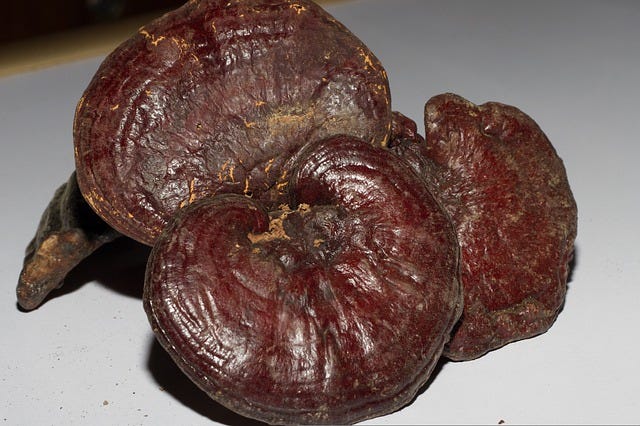
In a study attempting to find water extracts of herbal medicines targeting the SARS-Cov 2 virus 3CL protease and the RNA-dependent RNA polymerase, some of the active compounds identified in the screen were further tested in vivo, and it was found that extracts of Ganoderma lucidum (RF3) were effective in a challenge study using hamsters as disease model.
Jan, J. et al. 2021. “Identification of existing pharmaceuticals and herbal medicines as inhibitors of SARS-CoV-2 infection. Proc Natl Acad Sci U S A. 118(5):e2021579118.
This mushroom’s triterpene components have been demonstrated to exert inhibitory effects on the induction of Epstein-Barr virus early antigen (EBV-EA)
Iwatsuki K. et al. 2002. “Lucidenic acids P and Q, methyl lucidenate P, and other triterpenoids from the fungus Ganoderma lucidum and their inhibitory effects on Epstein-Barr virus activation,” J Nat Prod. 66(12):1582-5.
· Resveratrol
Reduces coronavirus inflammation.
Domi E. et al. 2022. “The Importance of Nutraceuticals in COVID-19: What's the Role of Resveratrol?” Molecules. 27(8):2376.
· Rice bran Arabinoxylan Compound (RBAC)
This immunomodulatory compound suppresses the degranulation of mast cells, which may be the, or a, factor in chronic inflammation in long-COVID. It also balances inflammatory cytokines as well as protects against oxidative stress. It has also shown benefits in chronic fatigue syndrome.
Ooi, S. 2021. “The Health-Promoting Properties and Clinical Applications of Rice Bran Arabinoxylan Modified with Shiitake Mushroom Enzyme-A Narrative Review,” Molecules. 26(9):2539.
· Selenium
Helps create antibodies. Deficiency allows greater virulence of RNA viruses like SARS-CoV-2. Protects the vascular endothelium. Helps to develop T-lymphocytes when there is lymphopenia, as is often the case in long-COVID. Is powerfully antioxidant.
Kieliszek, M. 2022. “Selenium in the Prevention of SARS-CoV-2 and Other Viruses,” Biol Trace Elem Res 19:1–8.
Avery J., Hoffmann, P. 2018. “Selenium, Selenoproteins, and Immunity,” Nutrients. 2018 10(9):1203.
· Self-Heal (Prunella vulgaris)
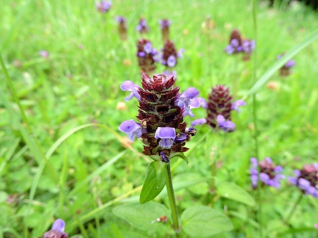
A scientific study found an extract of this herb to inhibit SARS CoV-2’s binding to cells. It has also demonstrated activity against Herpes-family viruses.
Ao, Z. et al. 2021. “Identification and evaluation of the inhibitory effect of Prunella vulgaris extract on SARS-coronavirus 2 virus entry,” PLoS One. 16(6):e0251649.
Chiu, L. et al. 2004. “A polysaccharide fraction from medicinal herb Prunella vulgaris downregulates the expression of herpes simplex virus antigen in Vero cells,” J. Ethnopharmacol. 93: 63–68.
Zhang, Y. et al. 2007. “Chemical properties, mode of action, and in vivo anti-herpes activities of a lignin-carbohydrate complex from Prunella vulgaris,” Antivir. Res. 75:242–249.
Moreover, this herb has been shown to manifest powerful immunomodulating/antihistamine/anti-inflammatory effects.
Fang, X. et al. 2005a. “Immune Modulatory Effects of Prunella vulgaris L.” Int J Mol Med, 15(3):491–96.
Fang, X, et al. 2005b. “Immune Modulatory Effects of Prunella vulgaris L. on Monocytes/Macrophages.” Int J MolMed. 16(6):1109–16.
Kim, S. et al. 2007. “Effects of Prunella vulgaris on Mast Cell-mediated Allergic Reaction and Inflammatory Cytokine Production.” Exp Biol Med (Maywood). 232(7):921–26.
· Turmeric (Curcuma longa)
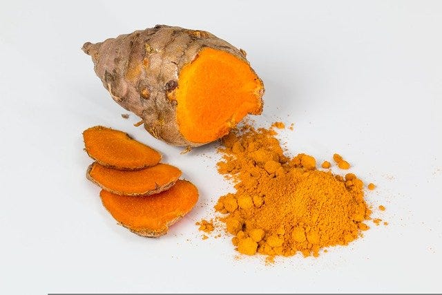
Scientific research has found turmeric to completely neutralize SARS-CoV-2 in vitro. Moreover, a scientific investigation of 36 extracts of 32 herbs belonging to 27 families found turmeric to be the most potent in terms of anti-EBV-EA activity—ten times more effective than passionflower, which was next in the order of activity.
Bormann, M. et al. 2021. “Turmeric root and its bioactive ingredient curcumin effectively neutralize SARS-CoV-2 in vitro,” Viruses. 13(10):1914
Kapadia, G. et al. 2002. “Inhibitory effect of herbal remedies on 12-O-tetradecanoylphorbol-13-acetate-promoted Epstein-Barr virus early antigen activation,” Pharmacol Res 45(3):213-20.
· Vitamin B-6
Helps the body make diamine oxidase (DAO), the enzyme that breaks done histamine (overactive in MCAC).
· Vitamin C
Positively influences lymphocyte development and function. Checks histamine. Stimulates interferon production from virus-infected cells, causing nearby cells to heighten their antiviral defenses. Restores the dysfunctional epithelial barrier of the lungs in respiratory infections. Many studies on the above functions. Also helps promote growth of protective bacterial populations in the gut. Intravenous vitamin C has been shown effective against both Epstein-Barr Virus and shingles.
Mikirova, N, Hunninghake, R. 2014. “Effect of high dose vitamin C on Epstein-Barr viral infection,” Med Sci Monit. 20:725-32.
· Vitamin D
An excellent immunomodulator and anti-inflammatory that has been shown in numerous studies to be vital for immune support during respiratory viruses and even for SARS-CoV-2 and Epstein-Barr Virus (EBV). Many published studies on the above. A dose that achieves a total vitamin-D serum level of 60-80 ng/mL needs to be used to achieve these effects. (This is usually at least 5,000 IU [= 125 mcg]/day.) Most people are deficient in vitamin D, according to wide-ranging nutritional surveys and research.
· Zinc
Supports T-lymphocytes, which are often deficient in long-COVID, as we have noted above. Supports membrane barrier integrity. Displays anti-inflammatory activity, and is involved in antibody production. It is also antithrombotic, antiviral, and antioxidant. Many studies on the above.
Wessels, I. et al. 2020. “The Potential Impact of Zinc Supplementation on COVID-19 Pathogenesis,” Front Immunol. 11:1712.
Support Adrenal Function & Mitochondria to Enhance Energy Levels
· N-acetyl Cysteine (NAC)
Prevents mitochondrial dysfunction from a variety of agents, but best used in combination with glycine.
Kumar, P. et al. 2021. “Glycine and N-acetylcysteine (GlyNAC) supplementation in older adults improves glutathione deficiency, oxidative stress, mitochondrial dysfunction, inflammation, insulin resistance, endothelial dysfunction, genotoxicity, muscle strength, and cognition: Results of a pilot clinical trial.” Clin Transl Med. 11(3):e372.
Shafie, B. et al. 2021. “N-acetylcysteine is more effective than ellagic acid in preventing acrolein induced dysfunction in mitochondria isolated from rat liver.” J Food Biochem. 45(7):e13775.
· Acetyl-L-carnitine
After long-COVID sufferers took a supplement containing vitamin C, acetyl-L-carnitine, B1, B6, folate, B12, D3, and olive polyphenols for 15 days, they recorded more improvement in energy and psychological status than did a control group.
Naureen, Z. 2021. “Proposal of a food supplement for the management of post-COVID syndrome,” Eur Rev Med Pharmacol Sci. 25(1 Suppl):67-73
· Adaptogens (Rhodiola, Eleuthero, Schizandra)
In a two-week-long, randomized, quadruple-blind, placebo-controlled clinical trial, supplementation with a fixed combination of adaptogens—rhodiola, eleuthero, and schizandra—decreased the duration of fatigue for one and two days, respectively, in 50% of 100 patients recruited for the study. The number of patients with lack of fatigue and pain symptoms was significantly less in the treatment group than in the placebo group on Days 9 and 11. The adaptogen mix also lowered the inflammatory cytokine IL-6 and creatinine (a marker of kidney failure) as against the placebo group
Karosanidze, I. et al. 2022. “Efficacy of Adaptogens in Patients with Long COVID-19: A Randomized, Quadruple-Blind, Placebo-Controlled Trial,” Pharmaceuticals (Basel). 15(3):345
· alpha-Lipoic acid
Supports mitochondrial function.
Liu, J. 2008. “The effects and mechanisms of mitochondrial nutrient alpha-lipoic acid on improving age-associated mitochondrial and cognitive dysfunction: An overview.” Neurochem. Res. 33:194–203.
Ong, S. et al. 2013. “The effect of alpha-lipoic acid on mitochondrial superoxide and glucocorticoid-induced hypertension,” Oxidative Med. Cell. Longev. 2013:517045.
· Coenzyme Q10
An interventional study has shown potential benefits of CoQ10 plus nicotinamide adenine dinucleotide (NAD) supplementation in patients with chronic fatigue syndrome (CFS), finding reduced maximum heart rate and perceptions of fatigue post-exercise testing. Typical dose: 200 mg/day, in divided doses
Castro-Marrero, J. et al. 2016. “Effect of coenzyme Q10 plus nicotinamide adenine dinucleotide supplementation on maximum heart rate after exercise testing in chronic fatigue syndrome—a randomized, controlled, double-blind trial,” Clin Nutr. 35(4):826–34.
· Probiotics
A clinical trial of hospitalized COVID-19 patients revealed a significant improvement in energy six months afterwards compared to those not given this therapy.
Santinelli, L. 2022. “Oral Bacteriotherapy Reduces the Occurrence of Chronic Fatigue in COVID-19 Patients,” Front Nutr. ;8:756177
· D-ribose
Has been shown to enhance energy and exercise recovery; reduces fatigue in CFS and in other persons with fatigue; improves cardiopulmonary function in exercise; and improves mental outlook. (Tastes delicious, too!)
Teitelbaum, J. et al. 2006. “The use of D-ribose in chronic fatigue syndrome and fibromyalgia: a pilot study,” J Altern Complement Med. 12:857–862.
Vijay N. et al. 2008. “D-ribose benefits heart failure patients.” J Med Food. 11(1):199-200.
MacCarter, D. 2009. “D-ribose aids advanced ischemic heart failure patients.” Int J Cardiol. 137(1):79-80.
· Selenium
Crucial to healthy mitochondrial function. Antioxidant.
Mehta, S. et al. 2012. “Selenium preserves mitochondrial function, stimulates mitochondrial biogenesis, and reduces infarct volume after focal cerebral ischemia,” BMC Neurosci. 13:79
· Vitamin B3 (Niacin)
A crucial source of energy production in the body—integral to all 3 stages of cellular respiration as a component of enzymes NAD & NADH and is active in electron transfer chains in mitochondria
· Vitamin B-complex
Improves energy levels through effects upon adrenal function, metabolism of macronutients/cellular respiration, energy production in the brain.
· Vitamin C
Intravenous vitamin C has been shown to improve fatigue in long-COVID patients.
Vollbracht, C., Kraft, K. 2021. “Feasibility of Vitamin C in the Treatment of Post Viral Fatigue with Focus on Long COVID, Based on a Systematic Review of IV Vitamin C on Fatigue,” Nutrients. 13(4):1154
Heal Damaged Body Systems/Organs
1. Cognitive Dysfunction / “Brain Fog” / Neuroinflammation
The “brain fog” described by sufferers of long-COVID is very likely related to neuroinflammation. Leaky gut (intestinal permeability) and dysbiosis have been shown to cause neuroinflammation by means of the bidirectional communication between the gut and the brain (the Gut-Brain-Microbiota Axis). Other neurological symptoms, resembling those experienced by sufferers of Parkinson’s disease, may also occur in Long-COVID sufferers.
· alpha-Lipoic Acid
Has a neuroprotective effect
Packer L, et al. 1997. “Neuroprotection by the metabolic antioxidant alpha-lipoic acid,” Free Radic Biol Med. 22(1-2):359-78.
· Perilla (Perilla frutescens) leaf extract
Perilla is rich in the flavonoid luteolin, a compound thought to be largely responsible for this herb’s widely appreciated antihistamine effects. May have benefits in brain fog.
Theoharides, T. et al. 2021. “Long-COVID syndrome-associated brain fog and chemofog: Luteolin to the rescue,” Biofactors. 47(2):232-241.
Marseglia, G. et al. 2019. “A polycentric, randomized, parallel-group, study on Lertal®, a multicomponent nutraceutical, as preventive treatment in children with allergic rhinoconjunctivitis: phase II,” Ital J Pediatr. 45(1):84.
· Probiotics
Support the intestinal barrier against permeability and reduce pathogenic organisms known to have adverse effects on the nervous system via bidirectional communication with the brain by means of the microbiota-gut-brain axis. Many studies.
· Turmeric (Curcuma longa)
An important antiinflammatory and neuroprotective agent. Many studies.
· Vitamin B1 (Thiamine)
Supports healthy nerve function—many studies. Note that alcohol, sugar, coffee and tea all interfere with vitamin B1 absorption and utilization. See the following:
· Vitamin D
An effective neuroprotectant, most likely even in COVID-19.
Quintero-Fabián, S. et al. 2022. “Vitamin D and its possible relationship to neuroprotection in COVID-19: evidence in the literature,” Curr Top Med Chem. 2022. Epub ahead of print.
2. Gustatory Sensory Loss (Anosmia/Aguesia)
Loss of the sense of smell, known as anosmia, and loss of the sense of taste (aguesia) are common features of long-COVID.
· Turmeric (Curcuma longa)
A fascinating case series narrates the speedy restoration of taste and smell in two long-COVID subjects following ingestion of a single 1000-mg dose of a turmeric supplement, standardized to 95% curcuminoids.
Chabot, A., Huntwork, M. 2021. “Turmeric as a Possible Treatment for COVID-19-Induced Anosmia and Ageusia.” Cureus. 13(9):e17829.
The following case report on reversing anosmia in a young, male COVID-19 patient was published in a medical journal. The following therapies were used:
· Thiamine (Vitamin B1), 100 mg/day,
· Vitamin B6, 100 mg/day
· Vitamin B12, 5 mg (= 5,000 mcg) day.
10-min. total/day of Inhalation Therapy, in divided sessions, was also used, implementing the following herbs/spices: cinnamon, cloves, and lavender. These were labeled for the patient so that the patient could make a point to remember what the smell of these spices was like and to associate that memory with trying to smell the herbs. In 16 days of treatment, sense of smell returned completely.
Pissurno, N. et al. 2020. “Anosmia in the course of COVID-19: A case report,” Medicine (Baltimore). 2020 Jul 31;99(31):e21280.
In a placebo-controlled clinical trial published in 2017, patients with post-viral anosmia used:
· Vitamin A liquid, applied topically (head back position) at a dose of 10,000 IU/day for 8 weeks. (This is typically only one drop with most liquid vitamin-A products available in the USA, wherein one drop = 10,000 IU.)
Results were: 37% of all post-infectious patients treated with vitamin A exhibited clinical improvement, whereas only 23% improved in controls.
Hummel, T. et al. 2017. “Intranasal vitamin A is beneficial in post-infectious olfactory loss,” Eur Arch Otorhinolaryngol. 274(7):2819-2825.
3. Cardiovascular & Respiratory Dysfunction
Patients with high concentrations of CRP and creatinine in the acute phase of Covid-19 are more prone to cardiac issues in long-COVID.
· N-acetyl Cysteine (NAC)
Supports overall respiratory function; thins mucus accumulation & promotes expectoration; helpful in shortness of breath; breaks down small blood clots called micro-emboli; increases low oxygen levels (as measured via an oximeter) and, in clinical studies, inhibits fibrous exudation in interstitial lung disease.
Zhou, N. et al. 2021. “The potential mechanism of N-acetylcysteine in treating COVID-19,” Curr Pharm Biotechnol. 22(12):1584-1590.
· Bromelain
This enzyme from pineapple is possessed of a potent fibrinolytic activity: it stimulates the conversion of plasminogen to plasmin, resulting in increased fibrinolysis by degrading fibrin (a protein involved in blood clotting) and it even inhibits the synthesis of fibrin. Many studies.
· Coenzyme Q10
Supports mitochondrial production of energy and left-ventricular health and displays antioxidant activity. Many studies.
· EPA/DHA (Omega-3 long-chain fatty acids)
Precursors of particles called resolvins D and E, which reduce proinflammatory mediators, thus decreasing inflammation. Many studies.
· Ginkgo biloba
Ginkgo has been shown to improve blood circulation and to support brain function at low oxygen levels, most likely by boosting oxygen saturation. Many studies. Several studies have even shown that it can help to prevent the common symptoms of altitude sickness.
· Hawthorn (Crataegus spp.)

This is a trophorestorative for the cardiovascular system. It has demonstrated anti-ischemic and endothelial-protective effects and has been shown to support myocardial health, strengthen the walls of blood vessels, and support a healthy blood pressure. Many studies.
· Olive (Olea europaea) leaf extract
Not only an excellent antiviral, as we’ve seen above, but also a vasodilator.
Zarzuelo, A. et al. 1991. “Vasodilator Effect of Olive Leaf,” Planta Medica 57(5):417-19.
· Quercetin
Has anti-thrombin effects, mitigating coagulation abnormalities.
Manjunath, S., Thimmulappa, R. 2022. “Antiviral, immunomodulatory, and anticoagulant effects of quercetin and its derivatives: Potential role in prevention and management of COVID-19.” J Pharm Anal. 12(1):29-34.
· Rhodiola (Rhodiola rosea)
SARS-CoV-2 infects immature red blood cells, which cannot carry oxygen, disallowing them to mature and to replace mature red blood cells that can carry oxygen but which only have a short life. Rhodiola has been shown to increase the efficiency of oxygen utilization and intracellular oxygen diffusion.
Ha, Z. et al. 2002. “[The effect of rhodiola and acetazolamide on the sleep architecture and blood oxygen saturation in men living at high altitude].” Zhonghua Jie He He Hu Xi Za Zhi. 25(9):527-30.
Ip, S. et al. 2001. “Association of free radicals and the tissue renin-angiotensin system: prospective effects of Rhodiola, a genus of Chinese herb, on hypoxia-induced pancreatic injury,” JOP. 2(1):16-25.
· Serrapeptase
This is an enzyme possibly capable of breaking down circulating spike proteins proven to be injurious to the cardiovascular system. Not to be taken concurrently with pharmaceutical anticoagulants.
· Vitamin C
Supports endothelial function
Ashor A. W. 2014. “Effect of vitamin C on endothelial function in health and disease: a systematic review and meta-analysis of randomised controlled trials.” Atherosclerosis. 235(1):9-20
Supplement Any Nutrient Deficiencies
Selenium
Vitamin C
Vitamin D
Zinc
Dietary Effects
Often helpful foods:
· High-sulfur foods (broccoli, cauliflower, kale, collard, turnip, radish, mustard greens, shallots, onions, Brussels sprouts, arugula, bok choy, and watercress.) These are all fibrinolytic, anti-inflammatory, and immunomodulatory. Moreover, sulfur nourishes mitochondria and helps synthesize glutathione.
https://www.sciencedirect.com/topics/medicine-and-dentistry/organosulfur-derivative
· Glutathione-supportive foods: Asparagus, avocado, green beans, spinach, watermelon, and cucumber.
· Carotene-rich foods (carrots, squash, spinach, etc.) or mixed carotenoids as a supplement
· Nitric-oxide increasing foods (nitric oxide relaxes blood vessels and makes it easier for a person to function in conditions of low oxygen): Beets, pomegranates, blueberries, dandelions
· Foods to increase the deficient probiotic Faecalibacterium prausnitzii: lingonberries; walnuts; chicory root; Jerusalem artichokes (sunchokes), red wine (or extracts thereof), and food sources of pectin (e.g., apples)
Unhelpful Foods:
· Processed foods; Sugar; High Omega-6 oils; Coffee
Dr. Bill Sardi noted that coffee consumption was the most common factor characterizing long-term COVID-19 treatment-resistant sufferers.